Amyvid
bb5a5043-0f51-11df-8a39-0800200c9a66
34391-3
HUMAN PRESCRIPTION DRUG LABEL
Drug Facts
Composition & Product
Identifiers & Packaging
Indications and Usage
AMYVID is indicated for positron emission tomography (PET) of the brain to estimate amyloid beta neuritic plaque density in adults with cognitive impairment for: Evaluation of Alzheimer's disease (AD) and other causes of cognitive decline Selection of patients who are indicated for amyloid beta-directed therapy as described in the prescribing information of the therapeutic products
Dosage and Administration
The recommended amount of radioactivity is 370 MBq (10 mCi) administered as a single intravenous bolus in a total volume of up to 10 mL. ( 2.2 ) Follow the injection with an intravenous flush of approximately 10 mL of 0.9% sodium chloride injection. ( 2.2 ) Obtain 10-minute PET images starting approximately 30 minutes to 50 minutes after drug administration. ( 2.3 ) See full prescribing information for image interpretation and radiation dosimetry. ( 2.4 , 2.5 )
Contraindications
None.
Warnings and Precautions
Risk of Image Misinterpretation and Other Errors: Image interpretation errors have been observed. ( 5.1 ) Radiation Risk: AMYVID contributes to a patient's long-term cumulative radiation exposure. Ensure safe drug handling to protect patients and health care providers from unintentional radiation exposure. Advise patients to hydrate before and after administration and to void frequently after administration. ( 2.1 , 2.2 , 5.2 )
Adverse Reactions
Most common adverse reactions (incidence ≥ 0.4%) were headache, musculoskeletal pain, increased blood pressure, nausea, fatigue, injection site reaction, anxiety, back pain, claustrophobia, dizziness, feeling cold, insomnia, and neck pain. ( 6.1 ) To report SUSPECTED ADVERSE REACTIONS, contact Eli Lilly and Company at 1-800-545-5979 or FDA at 1-800-FDA-1088 or www.fda.gov/medwatch .
Description
Indications and Usage ( 1 ) 6/2025 Dosage and Administration, Image Display and Interpretation ( 2.4 ) 6/2025
Medication Information
Warnings and Precautions
Risk of Image Misinterpretation and Other Errors: Image interpretation errors have been observed. ( 5.1 ) Radiation Risk: AMYVID contributes to a patient's long-term cumulative radiation exposure. Ensure safe drug handling to protect patients and health care providers from unintentional radiation exposure. Advise patients to hydrate before and after administration and to void frequently after administration. ( 2.1 , 2.2 , 5.2 )
Indications and Usage
AMYVID is indicated for positron emission tomography (PET) of the brain to estimate amyloid beta neuritic plaque density in adults with cognitive impairment for: Evaluation of Alzheimer's disease (AD) and other causes of cognitive decline Selection of patients who are indicated for amyloid beta-directed therapy as described in the prescribing information of the therapeutic products
Dosage and Administration
The recommended amount of radioactivity is 370 MBq (10 mCi) administered as a single intravenous bolus in a total volume of up to 10 mL. ( 2.2 ) Follow the injection with an intravenous flush of approximately 10 mL of 0.9% sodium chloride injection. ( 2.2 ) Obtain 10-minute PET images starting approximately 30 minutes to 50 minutes after drug administration. ( 2.3 ) See full prescribing information for image interpretation and radiation dosimetry. ( 2.4 , 2.5 )
Contraindications
None.
Adverse Reactions
Most common adverse reactions (incidence ≥ 0.4%) were headache, musculoskeletal pain, increased blood pressure, nausea, fatigue, injection site reaction, anxiety, back pain, claustrophobia, dizziness, feeling cold, insomnia, and neck pain. ( 6.1 ) To report SUSPECTED ADVERSE REACTIONS, contact Eli Lilly and Company at 1-800-545-5979 or FDA at 1-800-FDA-1088 or www.fda.gov/medwatch .
Description
Indications and Usage ( 1 ) 6/2025 Dosage and Administration, Image Display and Interpretation ( 2.4 ) 6/2025
Section 42229-5
Recommended Dosage
The recommended amount of radioactivity of AMYVID is 370 MBq (10 mCi) in a total volume of up to 10 mL, administered as a single intravenous bolus. The maximum mass dose is 50 mcg. Follow the injection with an intravenous flush of approximately 10 mL of 0.9% sodium chloride injection.
Section 43683-2
Section 51945-4
PACKAGE LABEL – Amyvid 50 mL PETNET Label
NDC Code 0002-1200-50
50 mL Multiple-Dose Vial
Sterile
Rx only
☢ CAUTION: RADIOACTIVE MATERIAL
AmyvidTM
Florbetapir F 18 Injection
____MBq (____mCi) in ____mL at ____:____ on ____
Batch No. ________________
For Intravenous Use.
Contains 0.1 to 19 micrograms of florbetapir, 4.5 mg sodium ascorbate USP and 0.1 mL dehydrated alcohol USP in 0.9% sodium chloride injection USP per milliliter of solution. Store at USP controlled room temperature 25ºC (77ºF); excursions permitted to 15ºC to 30ºC (59ºF to 86ºF). Stopre Amyvid within the original container or eqivalent radiation shielding.
Expires at ____:____ on ______________
Manufactured by PETNET Solutions, Inc. Knoxville, TN 37932 for Avid Radiopharmaceuticals, a wholly-owned subsidiary of Eli Lilly and Company, Philadelphia, PA 19104
10 Overdosage
In the event of administration of a radiation overdose with AMYVID, hydration and frequent urination should be encouraged to minimize radiation exposure to the patient.
8.4 Pediatric Use
The safety and effectiveness of AMYVID have not been established in pediatric patients.
8.5 Geriatric Use
Of the 496 patients in clinical studies of AMYVID, 307 patients were 65 years of age and older, while 203 patients were 75 years of age and older. No overall differences in safety or effectiveness were observed between patients 65 years of age and older and younger adult patients.
5.2 Radiation Risk
AMYVID contributes to a patient's overall long-term cumulative radiation exposure. Long-term cumulative radiation exposure is associated with an increased risk of cancer. Ensure safe drug handling to protect patients and health care providers from unintentional radiation exposure. Advise patients to hydrate before and after administration and to void frequently after administration [see Dosage and Administration (2.1, 2.2)].
4 Contraindications
None.
6 Adverse Reactions
Most common adverse reactions (incidence ≥ 0.4%) were headache, musculoskeletal pain, increased blood pressure, nausea, fatigue, injection site reaction, anxiety, back pain, claustrophobia, dizziness, feeling cold, insomnia, and neck pain. (6.1)
To report SUSPECTED ADVERSE REACTIONS, contact Eli Lilly and Company at 1-800-545-5979 or FDA at 1-800-FDA-1088 or www.fda.gov/medwatch .
12.2 Pharmacodynamics
Following intravenous injection, florbetapir F 18 diffuses across the human blood-brain barrier and produces a signal intensity detectable throughout the brain. Subsequently, cerebral perfusion decreases the brain florbetapir F 18 content, with differential retention of the drug in areas that contain amyloid beta aggregates compared to areas that lack the aggregates. The time-activity curves for florbetapir F 18 in the brain of subjects with positive scans show continual signal increases from time zero through 30 minutes post-administration, with stable values thereafter up to at least 90 minutes post-injection. Differences in signal intensity between brain regions showing specific and non-specific florbetapir F 18 uptake form the basis for the image interpretation method [see Dosage and Administration (2.4)].
The test-retest distribution of florbetapir F 18 was evaluated in 21 subjects (11 with probable AD and 10 healthy subjects) who underwent two administrations of florbetapir F 18 (followed by PET scans) separated by a time period of 2 to 30 days. Images were shown to maintain signal distribution reproducibility when evaluated qualitatively (by a reader blinded to image time points) as well as quantitatively using an automated assessment of standardized uptake value ratio (SUVR) in pre-specified brain regions. A comparison of a 10-minute versus a 20-minute image acquisition time showed no difference in the mean cortical to cerebellar SUVR results obtained.
12.3 Pharmacokinetics
Following intravenous administration of 370 MBq (10 mCi) of AMYVID in healthy subjects, florbetapir F 18 was distributed throughout the body with less than 5% of the injected F 18 radioactivity present in the blood by 20 minutes following administration, and less than 2% present by 45 minutes after administration. The F 18 in circulation during the 30-minute to 90-minute imaging window was principally in the form of polar florbetapir metabolites. Whole body scanning following the intravenous injection showed accumulation of radioactivity in the liver within 4 minutes post-injection, followed by elimination of the radioactivity primarily through the biliary/gastrointestinal tract. Some radioactivity is detected in the bladder. Essentially all radioactivity collected in the urine was present as polar metabolites of florbetapir F 18.
1 Indications and Usage
AMYVID is indicated for positron emission tomography (PET) of the brain to estimate amyloid beta neuritic plaque density in adults with cognitive impairment for:
- Evaluation of Alzheimer's disease (AD) and other causes of cognitive decline
- Selection of patients who are indicated for amyloid beta-directed therapy as described in the prescribing information of the therapeutic products
2.5 Radiation Dosimetry
Estimated radiation absorbed doses for adults from intravenous injection of AMYVID are shown in Table 1.
| ORGAN/TISSUE |
MEAN ABSORBED DOSE PER UNIT ADMINISTERED ACTIVITY
(microGy/MBq) |
| Adrenal | 14 |
| Brain | 10 |
| Breasts | 6 |
| Gallbladder Wall | 143 |
| Heart Wall | 13 |
| Kidneys | 14 |
| Liver | 64 |
| Lower Large Intestine Wall | 28 |
| Lungs | 9 |
| Muscle | 9 |
| Osteogenic Cells | 28 |
| Ovaries | 18 |
| Pancreas | 14 |
| Red Marrow | 14 |
| Skin | 6 |
| Small Intestine | 66 |
| Spleen | 9 |
| Stomach Wall | 12 |
| Testes | 7 |
| Thymus | 7 |
| Thyroid | 7 |
| Upper Large Intestine Wall | 74 |
| Urinary Bladder Wall | 27 |
| Uterus | 16 |
| Total Body | 12 |
| Effective Dose (microSv/MBq) | 19 |
The whole-body effective dose resulting from administration of 370 MBq (10 mCi) of AMYVID to an adult is estimated to be 7 mSv. When PET/CT is performed, exposure to radiation will increase by an amount dependent on the settings used in the CT acquisition.
12.1 Mechanism of Action
Florbetapir F 18 binds to amyloid beta plaques and the F 18 isotope produces a positron signal that is detected by a PET scanner. In in vitro binding studies using postmortem human brain homogenates containing amyloid beta plaques, the dissociation constant (Kd) for florbetapir was 3.7 ± 0.3 nM. The binding of florbetapir F 18 to amyloid beta aggregates was demonstrated in postmortem human brain sections using autoradiographic methods, thioflavin S and traditional silver staining correlation studies as well as immunohistochemistry (monoclonal antibody to amyloid beta) correlation studies. Florbetapir binding to tau protein and a battery of neuroreceptors was not detected in in vitro studies.
11.1 Drug Characteristics
AMYVID (florbetapir F 18 injection) is a radioactive diagnostic drug for intravenous use.
Chemically, florbetapir F 18 is (E)-4-(2-(6-(2-(2-(2[18F] fluoroethoxy)ethoxy)ethoxy)pyridine-3-yl)vinyl)-N-methylbenzamine. The molecular weight is 359 and the structural formula is:
AMYVID is a sterile, non-pyrogenic clear, colorless solution. Each mL contains 0.1 mcg to 19 mcg of florbetapir and 500 MBq to 1,900 MBq (13.5 mCi to 51 mCi) of florbetapir F 18 at EOS and the following inactive ingredients: 4.5 mg sodium ascorbate and 0.1 mL dehydrated alcohol in 0.9% sodium chloride injection. The pH of the solution is between 5.5 and 8.0.
5 Warnings and Precautions
- Risk of Image Misinterpretation and Other Errors: Image interpretation errors have been observed. (5.1)
- Radiation Risk: AMYVID contributes to a patient's long-term cumulative radiation exposure. Ensure safe drug handling to protect patients and health care providers from unintentional radiation exposure. Advise patients to hydrate before and after administration and to void frequently after administration. (2.1, 2.2, 5.2)
2 Dosage and Administration
- The recommended amount of radioactivity is 370 MBq (10 mCi) administered as a single intravenous bolus in a total volume of up to 10 mL. (2.2)
- Follow the injection with an intravenous flush of approximately 10 mL of 0.9% sodium chloride injection. (2.2)
- Obtain 10-minute PET images starting approximately 30 minutes to 50 minutes after drug administration. (2.3)
- See full prescribing information for image interpretation and radiation dosimetry. (2.4, 2.5)
3 Dosage Forms and Strengths
Injection: 500 MBq/mL to 1,900 MBq/mL (13.5 mCi/mL to 51 mCi/mL) of florbetapir F 18 in up to 100 mL volume at end of synthesis (EOS) as a clear, colorless solution in a multiple-dose vial.
8 Use in Specific Populations
Lactation: Temporarily discontinue breastfeeding. A lactating woman should pump and discard breast milk for 24 hours after AMYVID administration. (8.2)
6.1 Clinical Trials Experience
Because clinical trials are conducted under widely varying conditions, adverse reaction rates observed in the clinical trials of a drug cannot be directly compared to rates in the clinical trials of another drug and may not reflect the rates observed in clinical practice.
The safety of AMYVID was evaluated in 555 adult subjects who received AMYVID by intravenous injection in clinical trials. Table 2 shows adverse reactions reported in ≥ 0.4% of subjects from the clinical trials.
|
a Includes the terms blood pressure increased and hypertension. |
|
|
b Includes the terms injection site hemorrhage, injection site irritation, and injection site pain. |
|
|
c Includes the terms feeling cold and chills. |
|
| Adverse Reaction |
AMYVID
N=555 % |
| Headache | 1.8 |
| Musculoskeletal pain | 0.7 |
| Blood pressure increaseda | 0.7 |
| Nausea | 0.7 |
| Fatigue | 0.5 |
| Injection site reactionb | 0.5 |
| Anxiety | 0.4 |
| Back pain | 0.4 |
| Claustrophobia | 0.4 |
| Dizziness | 0.4 |
| Feeling coldc | 0.4 |
| Insomnia | 0.4 |
| Neck pain | 0.4 |
Adverse reactions that occurred in <0.4% of subjects included infusion site rash, dysgeusia, pruritus, urticaria, and flushing.
2.3 Image Acquisition Instructions
- Position the patient supine with the head positioned to center the brain, including the cerebellum, in the PET scanner field of view. Tape or other flexible head restraints may be employed to reduce head movement.
- Acquire 10-minute PET images starting 30 minutes to 50 minutes after AMYVID administration.
- Image reconstruction should include attenuation correction with resulting transaxial pixel sizes between 2 mm and 3 mm.
2.1 Radiation Safety Drug Handling
Handle AMYVID with appropriate safety measures to minimize radiation exposure during administration [see Warnings and Precautions (5.2)]. Use waterproof gloves and effective radiation shielding, including syringe shields when handling and administering AMYVID.
Radiopharmaceuticals, including AMYVID, should be used by or under the control of healthcare providers who are qualified by specific training and experience in the safe use and handling of radionuclides, and whose experience and training have been approved by the appropriate governmental agency authorized to license the use of radionuclides.
11.2 Nuclear Physical Characteristics
Fluorine-18 (F 18) decays by positron (β+) emission to oxygen-18 and has a physical half-life of 109.8 minutes. The principal photons useful for diagnostic imaging are the coincident pair of 511 keV gamma photons, resulting from the interaction of the emitted positron with an electron (Table 3).
| Radiation | Energy Level (keV) | Abundance (%) |
| Positron | 249.8 | 96.9 |
| Gamma | 511 | 193.5 |
The point source air-kerma coefficient for F 18 is 3.74E -17 Gy m2/(Bq s); this coefficient was formerly defined as the specific gamma-ray constant of 5.7 R/hr/mCi at 1 cm. The first half-value thickness of lead (Pb) for F 18 gamma rays is approximately 6 mm. The relative reduction of radiation emitted by F 18 that results from various thicknesses of lead shielding is shown in Table 4. The use of ~8 cm of Pb will decrease the radiation transmission (i.e., exposure) by a factor of about 10,000.
|
Shield Thickness
cm of Lead (Pb) |
Coefficient of Attenuation |
| 0.6 | 0.5 |
| 2 | 0.1 |
| 4 | 0.01 |
| 6 | 0.001 |
| 8 | 0.0001 |
For use in correcting for physical decay of this radionuclide, the fractions remaining at selected intervals after calibration are shown in Table 5.
| Minutes | Fraction Remaining |
| 0 | 1.00 |
| 15 | 0.909 |
| 30 | 0.826 |
| 60 | 0.683 |
| 110 | 0.500 |
| 220 | 0.250 |
| 440 | 0.060 |
5.1 Risk of Image Misinterpretation and Other Errors
Errors may occur in the estimation of brain amyloid beta neuritic plaque density during AMYVID image interpretation [see Clinical Studies (14)].
The use of clinical information in the interpretation of AMYVID images has not been evaluated and may lead to an inaccurate assessment. Extensive brain atrophy as well as motion artifacts that distort the image may limit the ability to distinguish gray and white matter on an AMYVID scan.
Perform image interpretation independently of the patient's clinical information. For cases where there is uncertainty as to the location of cortical signal, use co-registered anatomical imaging to improve localization of signal [see Dosage and Administration (2.4)].
13.1 Carcinogenesis, Mutagenesis, Impairment of Fertility
Animal studies to assess the carcinogenicity or reproductive toxicity potential of florbetapir F 18 have not been conducted.
14.1 Evaluation of Ad and Other Causes of Cognitive Decline
The effectiveness of AMYVID was evaluated in two single-arm clinical studies (i.e., Studies 1 and 2) in subjects with a range of cognitive function, including some terminally ill subjects who had agreed to participate in a postmortem brain donation program as well as healthy subjects. Subjects underwent an AMYVID injection and scan. The images were interpreted using a clinically applicable binary image interpretation method (negative or positive) by five independent readers who were blinded to all clinical information [see Dosage and Administration (2.3, 2.4)]. Image interpretations used co-registration with CT scans when PET scans were performed on dual PET-CT scanners. Before image interpretation, all readers underwent special training on image interpretation: in-person training or electronic media training.
The neuritic plaque density in both studies was determined using an algorithm in which microscopic measures of highest plaque density within a brain region were averaged to produce an estimate of global neuritic plaque density. The global neuritic plaque density was categorized in the same manner as that for a region (Table 6), where plaques were counted on slides with modified Bielschowsky silver stained tissue sections. To determine the agreement between the in vivo AMYVID image results and the postmortem amyloid beta neuritic plaque density, AMYVID results (negative or positive) were pre-specified to correspond to specific histopathology-derived plaque density scores, based upon a modification of the Consortium to Establish a Registry for Alzheimer's Disease (CERAD) criteria, which use neuritic plaque counts as a necessary pathological feature of AD.
| Histopathology Categorization | AMYVID PET Read | |
| Neuritic Plaque Counts | CERAD Score | |
| <1 | none | Negative |
| 1 - 5 | sparse | |
| 6 - 19 | moderate | Positive |
| 20+ | frequent |
Study 1 evaluated performance characteristics (sensitivity and specificity) in terminally ill subjects by comparing premortem AMYVID scans to a postmortem truth standard of amyloid beta neuritic plaque density. A total of 59 subjects underwent autopsy after being dosed with AMYVID and imaged. The mean age was 79 years (range 47 to 103 years), half were females, and most were White (93%). A total of 29 subjects had an AD clinical diagnosis, 13 had another type of dementing disorder, 5 had mild cognitive impairment (MCI), and 12 had no cognitive impairment. The time interval between the AMYVID scan and death was less than one year for 46 subjects and between one and two years for 13 subjects. At autopsy, the global brain neuritic plaque density category (CERAD score as in Table 6) was: frequent (n=30); moderate (n=9); sparse (n=5); and none (n=15).
Study 2 evaluated inter-reader and intra-reader reproducibility of image interpretation using images from 59 subjects with a truth standard (same subjects as in Study 1) and 92 subjects without a truth standard (52 subjects with MCI, 20 subjects with AD, and 20 healthy subjects). Intra-reader reproducibility was assessed with images from 33 subjects (22%). Among the 151 subjects, the mean age was 75 years (range 47 to 103 years), half were females, and most were White (93%).
Among all subjects who underwent autopsy within two years (n=59; 39 positive and 20 negative based on histopathology), the sensitivity using the majority interpretation of the readers trained in-person (Study 1) was 92% (95% CI: 78%, 98%) and specificity was 100% (95% CI: 80%, 100%). The median (and range) of correct, false negative, and false positive reads were 55 (45, 56), 3 (2, 12), 1 (0, 2), respectively, for in-person training; and were 51 (46 to 54), 7 (3 to 12), 1 (1 to 2), respectively, for electronic media training. Table 7 shows the inter-reader reproducibility results among readers who underwent electronic media training (Study 2) for various groups of subjects. Inter-reader reproducibility analyses for all images showed an overall Fleiss' kappa statistic of 0.83 (95% CI: 0.78, 0.88); the lower bound of the 95% CI exceeded the pre-specified success criterion of 0.58. Intra-reader reproducibility analyses showed that between the two readings for each of the 33 duplicate scans, one of the five readers had complete agreement for all 33 scans, two readers had discrepant reads for a single scan, one reader had discrepant reads for two scans, and another reader had discrepant reads for three scans.
|
a Readers who underwent electronic media training. |
|||||
|
b Shown is the median number of scans interpreted as positive across the five readers for each subgroup of subjects listed in the first column. |
|||||
| Subject Group by Cognitive Status and Truth Standard (TS) | Positive Scans, n b |
Kappa
(95% CI) |
Percent of Scans with Inter-Reader Agreement | ||
| 3 of 5 readers agreed | 4 of 5 readers agreed | 5 of 5 readers agreed | |||
| All subjects with a TS, n=59 | 33 | 0.75 (0.67, 0.83) |
14 | 10 | 76 |
| All subjects without a TS, n=92 | 33 | 0.88 (0.82, 0.94) |
2 | 11 | 87 |
| AD, n=49 (29 with TS; 20 no TS) |
38 | 0.67 (0.58, 0.76) |
10 | 14 | 76 |
| MCI, n=57 (5 with TS; 52 no TS) |
17 | 0.91 (0.83, 0.99) |
2 | 7 | 91 |
| Other non-AD dementia with TS, n=13 |
7 | 0.52 (0.35, 0.69) |
23 | 23 | 54 |
| Cognitively normal with TS, n=12 |
1 | 0.73 (0.55, 0.87) |
0 | 8 | 92 |
| Healthy subjects without TS, n=20 | 4 | 0.83 (0.69, 0.97) |
5 | 5 | 90 |
14.2 Selection of Patients Indicated for Amyloid Beta Directed Therapy
Refer to the Prescribing Information of amyloid beta-directed therapy for description of clinical trials in which the efficacy of amyloid beta PET for selecting patients has been established.
Brain amyloid beta PET scans have been used to assess reduction of plaque in some clinical trials of amyloid beta-directed therapies as also described in the prescribing information of the therapeutic products.
Structured Label Content
Section 42229-5 (42229-5)
Recommended Dosage
The recommended amount of radioactivity of AMYVID is 370 MBq (10 mCi) in a total volume of up to 10 mL, administered as a single intravenous bolus. The maximum mass dose is 50 mcg. Follow the injection with an intravenous flush of approximately 10 mL of 0.9% sodium chloride injection.
Section 43683-2 (43683-2)
Section 51945-4 (51945-4)
PACKAGE LABEL – Amyvid 50 mL PETNET Label
NDC Code 0002-1200-50
50 mL Multiple-Dose Vial
Sterile
Rx only
☢ CAUTION: RADIOACTIVE MATERIAL
AmyvidTM
Florbetapir F 18 Injection
____MBq (____mCi) in ____mL at ____:____ on ____
Batch No. ________________
For Intravenous Use.
Contains 0.1 to 19 micrograms of florbetapir, 4.5 mg sodium ascorbate USP and 0.1 mL dehydrated alcohol USP in 0.9% sodium chloride injection USP per milliliter of solution. Store at USP controlled room temperature 25ºC (77ºF); excursions permitted to 15ºC to 30ºC (59ºF to 86ºF). Stopre Amyvid within the original container or eqivalent radiation shielding.
Expires at ____:____ on ______________
Manufactured by PETNET Solutions, Inc. Knoxville, TN 37932 for Avid Radiopharmaceuticals, a wholly-owned subsidiary of Eli Lilly and Company, Philadelphia, PA 19104
10 Overdosage (10 OVERDOSAGE)
In the event of administration of a radiation overdose with AMYVID, hydration and frequent urination should be encouraged to minimize radiation exposure to the patient.
8.4 Pediatric Use
The safety and effectiveness of AMYVID have not been established in pediatric patients.
8.5 Geriatric Use
Of the 496 patients in clinical studies of AMYVID, 307 patients were 65 years of age and older, while 203 patients were 75 years of age and older. No overall differences in safety or effectiveness were observed between patients 65 years of age and older and younger adult patients.
5.2 Radiation Risk
AMYVID contributes to a patient's overall long-term cumulative radiation exposure. Long-term cumulative radiation exposure is associated with an increased risk of cancer. Ensure safe drug handling to protect patients and health care providers from unintentional radiation exposure. Advise patients to hydrate before and after administration and to void frequently after administration [see Dosage and Administration (2.1, 2.2)].
4 Contraindications (4 CONTRAINDICATIONS)
None.
6 Adverse Reactions (6 ADVERSE REACTIONS)
Most common adverse reactions (incidence ≥ 0.4%) were headache, musculoskeletal pain, increased blood pressure, nausea, fatigue, injection site reaction, anxiety, back pain, claustrophobia, dizziness, feeling cold, insomnia, and neck pain. (6.1)
To report SUSPECTED ADVERSE REACTIONS, contact Eli Lilly and Company at 1-800-545-5979 or FDA at 1-800-FDA-1088 or www.fda.gov/medwatch .
12.2 Pharmacodynamics
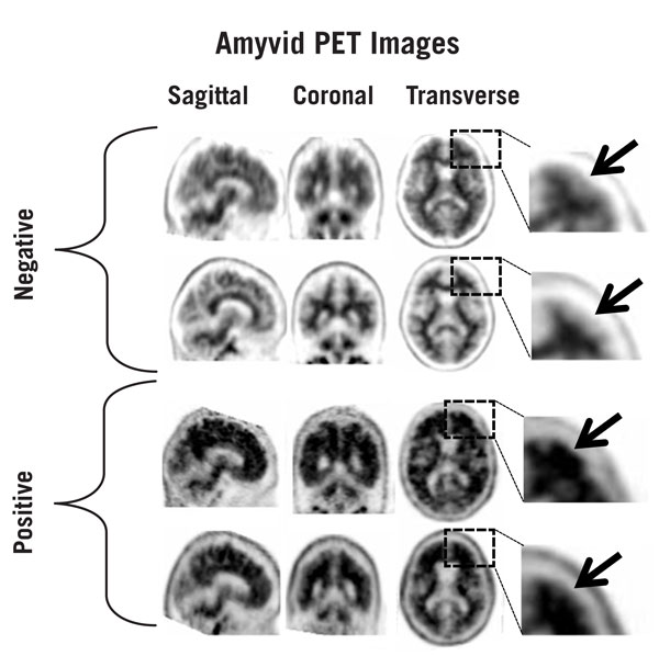
Following intravenous injection, florbetapir F 18 diffuses across the human blood-brain barrier and produces a signal intensity detectable throughout the brain. Subsequently, cerebral perfusion decreases the brain florbetapir F 18 content, with differential retention of the drug in areas that contain amyloid beta aggregates compared to areas that lack the aggregates. The time-activity curves for florbetapir F 18 in the brain of subjects with positive scans show continual signal increases from time zero through 30 minutes post-administration, with stable values thereafter up to at least 90 minutes post-injection. Differences in signal intensity between brain regions showing specific and non-specific florbetapir F 18 uptake form the basis for the image interpretation method [see Dosage and Administration (2.4)].
The test-retest distribution of florbetapir F 18 was evaluated in 21 subjects (11 with probable AD and 10 healthy subjects) who underwent two administrations of florbetapir F 18 (followed by PET scans) separated by a time period of 2 to 30 days. Images were shown to maintain signal distribution reproducibility when evaluated qualitatively (by a reader blinded to image time points) as well as quantitatively using an automated assessment of standardized uptake value ratio (SUVR) in pre-specified brain regions. A comparison of a 10-minute versus a 20-minute image acquisition time showed no difference in the mean cortical to cerebellar SUVR results obtained.
12.3 Pharmacokinetics
Following intravenous administration of 370 MBq (10 mCi) of AMYVID in healthy subjects, florbetapir F 18 was distributed throughout the body with less than 5% of the injected F 18 radioactivity present in the blood by 20 minutes following administration, and less than 2% present by 45 minutes after administration. The F 18 in circulation during the 30-minute to 90-minute imaging window was principally in the form of polar florbetapir metabolites. Whole body scanning following the intravenous injection showed accumulation of radioactivity in the liver within 4 minutes post-injection, followed by elimination of the radioactivity primarily through the biliary/gastrointestinal tract. Some radioactivity is detected in the bladder. Essentially all radioactivity collected in the urine was present as polar metabolites of florbetapir F 18.
1 Indications and Usage (1 INDICATIONS AND USAGE)
AMYVID is indicated for positron emission tomography (PET) of the brain to estimate amyloid beta neuritic plaque density in adults with cognitive impairment for:
- Evaluation of Alzheimer's disease (AD) and other causes of cognitive decline
- Selection of patients who are indicated for amyloid beta-directed therapy as described in the prescribing information of the therapeutic products
2.5 Radiation Dosimetry
Estimated radiation absorbed doses for adults from intravenous injection of AMYVID are shown in Table 1.
| ORGAN/TISSUE |
MEAN ABSORBED DOSE PER UNIT ADMINISTERED ACTIVITY
(microGy/MBq) |
| Adrenal | 14 |
| Brain | 10 |
| Breasts | 6 |
| Gallbladder Wall | 143 |
| Heart Wall | 13 |
| Kidneys | 14 |
| Liver | 64 |
| Lower Large Intestine Wall | 28 |
| Lungs | 9 |
| Muscle | 9 |
| Osteogenic Cells | 28 |
| Ovaries | 18 |
| Pancreas | 14 |
| Red Marrow | 14 |
| Skin | 6 |
| Small Intestine | 66 |
| Spleen | 9 |
| Stomach Wall | 12 |
| Testes | 7 |
| Thymus | 7 |
| Thyroid | 7 |
| Upper Large Intestine Wall | 74 |
| Urinary Bladder Wall | 27 |
| Uterus | 16 |
| Total Body | 12 |
| Effective Dose (microSv/MBq) | 19 |
The whole-body effective dose resulting from administration of 370 MBq (10 mCi) of AMYVID to an adult is estimated to be 7 mSv. When PET/CT is performed, exposure to radiation will increase by an amount dependent on the settings used in the CT acquisition.
12.1 Mechanism of Action
Florbetapir F 18 binds to amyloid beta plaques and the F 18 isotope produces a positron signal that is detected by a PET scanner. In in vitro binding studies using postmortem human brain homogenates containing amyloid beta plaques, the dissociation constant (Kd) for florbetapir was 3.7 ± 0.3 nM. The binding of florbetapir F 18 to amyloid beta aggregates was demonstrated in postmortem human brain sections using autoradiographic methods, thioflavin S and traditional silver staining correlation studies as well as immunohistochemistry (monoclonal antibody to amyloid beta) correlation studies. Florbetapir binding to tau protein and a battery of neuroreceptors was not detected in in vitro studies.
11.1 Drug Characteristics
AMYVID (florbetapir F 18 injection) is a radioactive diagnostic drug for intravenous use.
Chemically, florbetapir F 18 is (E)-4-(2-(6-(2-(2-(2[18F] fluoroethoxy)ethoxy)ethoxy)pyridine-3-yl)vinyl)-N-methylbenzamine. The molecular weight is 359 and the structural formula is:
AMYVID is a sterile, non-pyrogenic clear, colorless solution. Each mL contains 0.1 mcg to 19 mcg of florbetapir and 500 MBq to 1,900 MBq (13.5 mCi to 51 mCi) of florbetapir F 18 at EOS and the following inactive ingredients: 4.5 mg sodium ascorbate and 0.1 mL dehydrated alcohol in 0.9% sodium chloride injection. The pH of the solution is between 5.5 and 8.0.
5 Warnings and Precautions (5 WARNINGS AND PRECAUTIONS)
- Risk of Image Misinterpretation and Other Errors: Image interpretation errors have been observed. (5.1)
- Radiation Risk: AMYVID contributes to a patient's long-term cumulative radiation exposure. Ensure safe drug handling to protect patients and health care providers from unintentional radiation exposure. Advise patients to hydrate before and after administration and to void frequently after administration. (2.1, 2.2, 5.2)
2 Dosage and Administration (2 DOSAGE AND ADMINISTRATION)
- The recommended amount of radioactivity is 370 MBq (10 mCi) administered as a single intravenous bolus in a total volume of up to 10 mL. (2.2)
- Follow the injection with an intravenous flush of approximately 10 mL of 0.9% sodium chloride injection. (2.2)
- Obtain 10-minute PET images starting approximately 30 minutes to 50 minutes after drug administration. (2.3)
- See full prescribing information for image interpretation and radiation dosimetry. (2.4, 2.5)
3 Dosage Forms and Strengths (3 DOSAGE FORMS AND STRENGTHS)
Injection: 500 MBq/mL to 1,900 MBq/mL (13.5 mCi/mL to 51 mCi/mL) of florbetapir F 18 in up to 100 mL volume at end of synthesis (EOS) as a clear, colorless solution in a multiple-dose vial.
8 Use in Specific Populations (8 USE IN SPECIFIC POPULATIONS)
Lactation: Temporarily discontinue breastfeeding. A lactating woman should pump and discard breast milk for 24 hours after AMYVID administration. (8.2)
6.1 Clinical Trials Experience
Because clinical trials are conducted under widely varying conditions, adverse reaction rates observed in the clinical trials of a drug cannot be directly compared to rates in the clinical trials of another drug and may not reflect the rates observed in clinical practice.
The safety of AMYVID was evaluated in 555 adult subjects who received AMYVID by intravenous injection in clinical trials. Table 2 shows adverse reactions reported in ≥ 0.4% of subjects from the clinical trials.
|
a Includes the terms blood pressure increased and hypertension. |
|
|
b Includes the terms injection site hemorrhage, injection site irritation, and injection site pain. |
|
|
c Includes the terms feeling cold and chills. |
|
| Adverse Reaction |
AMYVID
N=555 % |
| Headache | 1.8 |
| Musculoskeletal pain | 0.7 |
| Blood pressure increaseda | 0.7 |
| Nausea | 0.7 |
| Fatigue | 0.5 |
| Injection site reactionb | 0.5 |
| Anxiety | 0.4 |
| Back pain | 0.4 |
| Claustrophobia | 0.4 |
| Dizziness | 0.4 |
| Feeling coldc | 0.4 |
| Insomnia | 0.4 |
| Neck pain | 0.4 |
Adverse reactions that occurred in <0.4% of subjects included infusion site rash, dysgeusia, pruritus, urticaria, and flushing.
2.3 Image Acquisition Instructions
- Position the patient supine with the head positioned to center the brain, including the cerebellum, in the PET scanner field of view. Tape or other flexible head restraints may be employed to reduce head movement.
- Acquire 10-minute PET images starting 30 minutes to 50 minutes after AMYVID administration.
- Image reconstruction should include attenuation correction with resulting transaxial pixel sizes between 2 mm and 3 mm.
2.1 Radiation Safety Drug Handling (2.1 Radiation Safety - Drug Handling)
Handle AMYVID with appropriate safety measures to minimize radiation exposure during administration [see Warnings and Precautions (5.2)]. Use waterproof gloves and effective radiation shielding, including syringe shields when handling and administering AMYVID.
Radiopharmaceuticals, including AMYVID, should be used by or under the control of healthcare providers who are qualified by specific training and experience in the safe use and handling of radionuclides, and whose experience and training have been approved by the appropriate governmental agency authorized to license the use of radionuclides.
11.2 Nuclear Physical Characteristics
Fluorine-18 (F 18) decays by positron (β+) emission to oxygen-18 and has a physical half-life of 109.8 minutes. The principal photons useful for diagnostic imaging are the coincident pair of 511 keV gamma photons, resulting from the interaction of the emitted positron with an electron (Table 3).
| Radiation | Energy Level (keV) | Abundance (%) |
| Positron | 249.8 | 96.9 |
| Gamma | 511 | 193.5 |
The point source air-kerma coefficient for F 18 is 3.74E -17 Gy m2/(Bq s); this coefficient was formerly defined as the specific gamma-ray constant of 5.7 R/hr/mCi at 1 cm. The first half-value thickness of lead (Pb) for F 18 gamma rays is approximately 6 mm. The relative reduction of radiation emitted by F 18 that results from various thicknesses of lead shielding is shown in Table 4. The use of ~8 cm of Pb will decrease the radiation transmission (i.e., exposure) by a factor of about 10,000.
|
Shield Thickness
cm of Lead (Pb) |
Coefficient of Attenuation |
| 0.6 | 0.5 |
| 2 | 0.1 |
| 4 | 0.01 |
| 6 | 0.001 |
| 8 | 0.0001 |
For use in correcting for physical decay of this radionuclide, the fractions remaining at selected intervals after calibration are shown in Table 5.
| Minutes | Fraction Remaining |
| 0 | 1.00 |
| 15 | 0.909 |
| 30 | 0.826 |
| 60 | 0.683 |
| 110 | 0.500 |
| 220 | 0.250 |
| 440 | 0.060 |
5.1 Risk of Image Misinterpretation and Other Errors
Errors may occur in the estimation of brain amyloid beta neuritic plaque density during AMYVID image interpretation [see Clinical Studies (14)].
The use of clinical information in the interpretation of AMYVID images has not been evaluated and may lead to an inaccurate assessment. Extensive brain atrophy as well as motion artifacts that distort the image may limit the ability to distinguish gray and white matter on an AMYVID scan.
Perform image interpretation independently of the patient's clinical information. For cases where there is uncertainty as to the location of cortical signal, use co-registered anatomical imaging to improve localization of signal [see Dosage and Administration (2.4)].
13.1 Carcinogenesis, Mutagenesis, Impairment of Fertility
Animal studies to assess the carcinogenicity or reproductive toxicity potential of florbetapir F 18 have not been conducted.
14.1 Evaluation of Ad and Other Causes of Cognitive Decline (14.1 Evaluation of AD and Other Causes of Cognitive Decline)
The effectiveness of AMYVID was evaluated in two single-arm clinical studies (i.e., Studies 1 and 2) in subjects with a range of cognitive function, including some terminally ill subjects who had agreed to participate in a postmortem brain donation program as well as healthy subjects. Subjects underwent an AMYVID injection and scan. The images were interpreted using a clinically applicable binary image interpretation method (negative or positive) by five independent readers who were blinded to all clinical information [see Dosage and Administration (2.3, 2.4)]. Image interpretations used co-registration with CT scans when PET scans were performed on dual PET-CT scanners. Before image interpretation, all readers underwent special training on image interpretation: in-person training or electronic media training.
The neuritic plaque density in both studies was determined using an algorithm in which microscopic measures of highest plaque density within a brain region were averaged to produce an estimate of global neuritic plaque density. The global neuritic plaque density was categorized in the same manner as that for a region (Table 6), where plaques were counted on slides with modified Bielschowsky silver stained tissue sections. To determine the agreement between the in vivo AMYVID image results and the postmortem amyloid beta neuritic plaque density, AMYVID results (negative or positive) were pre-specified to correspond to specific histopathology-derived plaque density scores, based upon a modification of the Consortium to Establish a Registry for Alzheimer's Disease (CERAD) criteria, which use neuritic plaque counts as a necessary pathological feature of AD.
| Histopathology Categorization | AMYVID PET Read | |
| Neuritic Plaque Counts | CERAD Score | |
| <1 | none | Negative |
| 1 - 5 | sparse | |
| 6 - 19 | moderate | Positive |
| 20+ | frequent |
Study 1 evaluated performance characteristics (sensitivity and specificity) in terminally ill subjects by comparing premortem AMYVID scans to a postmortem truth standard of amyloid beta neuritic plaque density. A total of 59 subjects underwent autopsy after being dosed with AMYVID and imaged. The mean age was 79 years (range 47 to 103 years), half were females, and most were White (93%). A total of 29 subjects had an AD clinical diagnosis, 13 had another type of dementing disorder, 5 had mild cognitive impairment (MCI), and 12 had no cognitive impairment. The time interval between the AMYVID scan and death was less than one year for 46 subjects and between one and two years for 13 subjects. At autopsy, the global brain neuritic plaque density category (CERAD score as in Table 6) was: frequent (n=30); moderate (n=9); sparse (n=5); and none (n=15).
Study 2 evaluated inter-reader and intra-reader reproducibility of image interpretation using images from 59 subjects with a truth standard (same subjects as in Study 1) and 92 subjects without a truth standard (52 subjects with MCI, 20 subjects with AD, and 20 healthy subjects). Intra-reader reproducibility was assessed with images from 33 subjects (22%). Among the 151 subjects, the mean age was 75 years (range 47 to 103 years), half were females, and most were White (93%).
Among all subjects who underwent autopsy within two years (n=59; 39 positive and 20 negative based on histopathology), the sensitivity using the majority interpretation of the readers trained in-person (Study 1) was 92% (95% CI: 78%, 98%) and specificity was 100% (95% CI: 80%, 100%). The median (and range) of correct, false negative, and false positive reads were 55 (45, 56), 3 (2, 12), 1 (0, 2), respectively, for in-person training; and were 51 (46 to 54), 7 (3 to 12), 1 (1 to 2), respectively, for electronic media training. Table 7 shows the inter-reader reproducibility results among readers who underwent electronic media training (Study 2) for various groups of subjects. Inter-reader reproducibility analyses for all images showed an overall Fleiss' kappa statistic of 0.83 (95% CI: 0.78, 0.88); the lower bound of the 95% CI exceeded the pre-specified success criterion of 0.58. Intra-reader reproducibility analyses showed that between the two readings for each of the 33 duplicate scans, one of the five readers had complete agreement for all 33 scans, two readers had discrepant reads for a single scan, one reader had discrepant reads for two scans, and another reader had discrepant reads for three scans.
|
a Readers who underwent electronic media training. |
|||||
|
b Shown is the median number of scans interpreted as positive across the five readers for each subgroup of subjects listed in the first column. |
|||||
| Subject Group by Cognitive Status and Truth Standard (TS) | Positive Scans, n b |
Kappa
(95% CI) |
Percent of Scans with Inter-Reader Agreement | ||
| 3 of 5 readers agreed | 4 of 5 readers agreed | 5 of 5 readers agreed | |||
| All subjects with a TS, n=59 | 33 | 0.75 (0.67, 0.83) |
14 | 10 | 76 |
| All subjects without a TS, n=92 | 33 | 0.88 (0.82, 0.94) |
2 | 11 | 87 |
| AD, n=49 (29 with TS; 20 no TS) |
38 | 0.67 (0.58, 0.76) |
10 | 14 | 76 |
| MCI, n=57 (5 with TS; 52 no TS) |
17 | 0.91 (0.83, 0.99) |
2 | 7 | 91 |
| Other non-AD dementia with TS, n=13 |
7 | 0.52 (0.35, 0.69) |
23 | 23 | 54 |
| Cognitively normal with TS, n=12 |
1 | 0.73 (0.55, 0.87) |
0 | 8 | 92 |
| Healthy subjects without TS, n=20 | 4 | 0.83 (0.69, 0.97) |
5 | 5 | 90 |
14.2 Selection of Patients Indicated for Amyloid Beta Directed Therapy (14.2 Selection of Patients Indicated for Amyloid Beta-Directed Therapy)
Refer to the Prescribing Information of amyloid beta-directed therapy for description of clinical trials in which the efficacy of amyloid beta PET for selecting patients has been established.
Brain amyloid beta PET scans have been used to assess reduction of plaque in some clinical trials of amyloid beta-directed therapies as also described in the prescribing information of the therapeutic products.
Advanced Ingredient Data
Raw Label Data
All Sections (JSON)
Additional Information
Back to search View SPL set listing Open on DailyMed ↗
Source: dailymed · Ingested: 2026-02-15T11:53:13.695596 · Updated: 2026-03-14T22:45:57.416825